- REAGENT SERVICES Hot!
-
PRODUCTS
-
 Most Popular Reagents
-
 Instruments
-
 Antibodies
-
ELISA Kits
-
 Protein Electrophoresis and Blotting
-
Protein and Antibody Purification
-
Recombinant Proteins
-
 Molecular Biology
-
 Stable Cell Lines
-
 Cell Isolation and Activation
-
 IVD Raw Materials
-
 Therapy Applications
-
 Resources
-
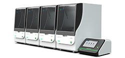
![AmMag™ Quatro Automated Plasmid Purification]() AmMag™ Quatro automated plasmid purification
AmMag™ Quatro automated plasmid purification
-
![Anti-Camelid VHH]() MonoRab™ Anti-VHH Antibodies
MonoRab™ Anti-VHH Antibodies
-
![ELISA Kits]() ELISA Kits
ELISA Kits
-
![Precast Gels]() SurePAGE™ Precast Gels
SurePAGE™ Precast Gels
-
![Quatro ProAb Automated Protein and Antibody Purification System]() AmMag™ Quatro ProAb Automated Protein and Antibody Purification System
AmMag™ Quatro ProAb Automated Protein and Antibody Purification System
-
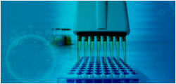
![Target Proteins]() Target Proteins
Target Proteins
-
![AmMag™ Quatro Automated Plasmid Purification]() AmMag™ Quatro automated plasmid purification
AmMag™ Quatro automated plasmid purification
-
![Stable Cell Lines]() Stable Cell Lines
Stable Cell Lines
-
![Cell Isolation and Activation]() Cell Isolation and Activation
Cell Isolation and Activation
-
![Quick
Order]() Quick Order
Quick Order
-
![Quick
Order]() Quick Order
Quick Order
- APPLICATIONS
- RESOURCES
- ABOUT US


List by Alphabet: A B C D E F G H I J K L M N O P Q R S T U V W X Y Z
antibody staining
In the realm of cellular and molecular biology, antibody staining, also known as immunohistochemistry (IHC), stands as a powerful technique, allowing researchers to delve into the intricate details of cellular components. This comprehensive guide explores the fundamentals of antibody staining, its applications, and the crucial role it plays in unraveling the mysteries of biological systems.
Understanding Antibody Staining: A Brief Overview
Antibody staining is a sophisticated laboratory technique used to visualize specific proteins or antigens within cells or tissues. This process relies on the use of antibodies—proteins produced by the immune system—that exhibit a remarkable ability to bind selectively to target molecules.
The Dance of Molecules: Key Steps in Antibody Staining
1. Fixation: The journey begins with the fixation of tissues or cells to preserve their structural integrity.
2. Permeabilization: Cell membranes may be permeabilized to allow antibodies to penetrate and interact with intracellular targets.
3. Blocking: To minimize nonspecific binding, researchers employ blocking agents to neutralize unoccupied binding sites.
4. Primary Antibody Incubation: The star of the show, the primary antibody, is introduced. This antibody, specific to the target antigen, binds with precision.
5. Washing: Excess primary antibody is washed away, reducing background noise and enhancing specificity.
6. Secondary Antibody Incubation: A secondary antibody, conjugated to a detectable label like a fluorescent dye or enzyme, amplifies the signal.
7. Washing (Again): Thorough washing ensures removal of excess secondary antibody, refining the signal-to-noise ratio.
8. Detection: The stained cells or tissues are ready for their close-up. Whether under a microscope or through advanced imaging techniques, researchers can now visualize the distribution and abundance of specific proteins.
Applications Across the Scientific Landscape
Research Advancements:
Antibody staining is a cornerstone in biological research, enabling scientists to investigate cellular processes, identify biomarkers, and understand the mechanisms underlying diseases such as cancer and neurodegenerative disorders.
Diagnostic Precision:
In clinical settings, antibody staining aids in disease diagnosis and prognosis. Pathologists use this technique to detect specific antigens, contributing to more accurate disease classification and treatment planning.
Therapeutic Developments:
The technique plays a pivotal role in the development of targeted therapies. By visualizing the presence and distribution of specific proteins, researchers can tailor therapeutic interventions for maximum effectiveness.
Looking Ahead: Innovations in Antibody Staining Techniques
As technology advances, so does antibody staining. Emerging methods, including multiplex staining and super-resolution microscopy, push the boundaries of what is possible, allowing researchers to simultaneously visualize multiple targets and achieve higher resolution images.
Conclusion: Unveiling the Microscopic Universe
Antibody staining is not merely a laboratory technique; it is a key that unlocks the microscopic universe, revealing the secrets of cellular function and dysfunction. As researchers continue to refine and expand antibody staining methodologies, the potential for groundbreaking discoveries in medicine and biology remains vast.
In the ever-evolving landscape of scientific exploration, antibody staining stands as a beacon, guiding researchers toward a deeper understanding of the complex tapestry that is life at the cellular level.
- Tags:
- Antibody
Related Biology Tools
-
GenSmart™ Codon Optimization
GenSmart Optimization is a free online tool for performing codon optimization to improve gene expression. GenScript's patented algorithms are integrated into the tool to optimize the computing capability of high-performance sequence generation.
-
DNA Construct Design Tool
GenSmart™ Design is a free online DNA construct design tool developed by GenScript. GenSmart™ Design has two design modules, the Create Construct module for individual plasmid design and the Create Library module for DNA library design.
-
Codon Frequency Tables
This online tool shows commonly used genetic codon frequency table in expression host organisms including Escherichia coli and other common host organisms.
Service and Products

Antibody Production Services
Comprehensive custom antibody production services including monoclonal and polyclonal antibody production services with industry leading turnaround times.

Polyclonal Antibody Services
The fastest custom polyclonal antibody production services in the industry with unmatched guarantees.

Custom Monoclonal Antibody Development Services
Developing custom monoclonal antibodies with standard or fully custom protocols including everything from antigen synthesis to antibody scale-up options.

Phospho-Specific Antibody Services
Guaranteed ELISA titer of ≥ 1:64,000 and < 10% cross reactivity with non-phospho peptide.

Immunoassay Development Services
Our custom immunoassay development service offering comprehensive high-throughput assay development with fast turnaround and high efficiency.

Large-scale Antibody Manufacturing
Our large-scale antibody manufacturing services offer polyclonal and monoclonal antibody production of gram quantities for industrial antibody yields.

COVID-19 Reagent Antibodies
On demand SARS-CoV-2 control mAbs ideal for diagnostics, therapeutic, and vaccine development.
-
Top Search
-
Hot Glossary
-
Antibody
If you know of any terms that have been omitted from this glossary that you feel would be useful to include, please send detail to the Editorial Office at GenScript: website@genscript.com
If your term is adopted, we will send 1,000 EzCoupon points to your GenScript account. -