Resources » Weekly Scientific Insight » Optimizing Sensitivity in Antibody-Based Immunoassays: Key Strategies for Reliable Diagnostics
Editor: Amanda Grimm
Sept 12, 2024
In the world of in vitro diagnostics, the sensitivity of an assay can often be the difference between early detection and a missed diagnosis 1. Sensitivity refers to an assay's ability to detect even the smallest amounts of a target biomarker, making it a critical parameter in developing antibody-based immunoassays. Whether you're working on detecting infectious diseases 2, cancer markers, or other low-abundance biomarkers, optimizing sensitivity is essential to achieving reliable and clinically relevant results.
But let’s dive into why sensitivity matters! In clinical settings, a highly sensitive immunoassay can identify biomarkers at the earliest stages of a disease, allowing for timely intervention and improving patient outcomes. Conversely, a lack of sensitivity can result in false negatives, potentially delaying critical treatment. As the demand for precision diagnostics continues to rise, the need for ultra-sensitive assays has never been greater.
This article will dive into the key factors that influence assay sensitivity and explore practical strategies to optimize detection limits in antibody-based immunoassays. From selecting high-affinity antibodies to minimizing background noise and enhancing signal detection, we'll cover everything you need to know to maximize the performance of your diagnostic assays.
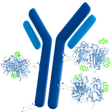
Positive and negative antibody-based immunoassay.
When it comes to developing a high-performing antibody-based immunoassay, the choice of reagents particularly the antibody, is crucial3. Antibodies and antigens lead the IVD raw materials market4. Two key properties that directly impact the sensitivity of your assay are antibody affinity and specificity. Understanding and optimizing these characteristics are fundamental to ensuring the assay accurately detects the target biomarker, even at very low concentrations.
Antibody Affinity: The Strength of Binding
Antibody affinity refers to the strength of the interaction between an antibody and its specific antigen. High-affinity antibodies bind more tightly to their target, which is critical to detect low-abundance biomarkers. This strong binding ensures that even minute quantities of the antigen are captured effectively, leading to a detectable signal 5. In contrast, low-affinity antibodies may dissociate from the antigen more easily, reducing the likelihood of detection and potentially leading to false negatives.
Optimizing affinity is particularly important when dealing with complex biological samples, where the target antigen may be present in very low concentrations amidst a plethora of other molecules. High-affinity antibodies help distinguish the target from background noise, which improves the overall sensitivity of the assay.
Antibody Specificity: Precision in Target Recognition
Antibody specificity defines how precisely an antibody recognizes and binds to its intended target without cross-reacting with other molecules.
In antibody-based immunoassays, specificity is critical to ensure that the detected signal truly corresponds to the target antigen and not to unrelated proteins or other substances present in the sample. Cross-reactivity can significantly compromise the assay's accuracy, leading to misleading false-positive results that could have serious implications in a clinical setting.
Balancing Affinity and Specificity
While both affinity and specificity are vital, achieving a balance between the two is key. An antibody with extremely high affinity but poor specificity could bind to non-target molecules with similar structures, leading to false positives. Conversely, an antibody with high specificity but low affinity might miss low-concentration targets, resulting in false negatives.
To optimize both affinity and specificity, the antibody selection process often involves screening a large library of candidates to identify those that meet the desired criteria. Techniques such as phage display, hybridoma technology, and recombinant antibody engineering are commonly used to develop antibodies with the ideal balance of these properties.
Signal amplification is an important consideration for immunoassay developers, that contributes to the detection of the smallest amount of target molecules. In antibody-based immunoassays, leveraging the right amplification techniques dramatically enhances the assay's ability to detect low-abundance biomarkers, that translates into improved diagnostic accuracy and earlier disease detection.
Signal amplification involves enhancing the signal generated by the binding of an antibody to its target antigen, making it easier to detect even when the antigen is present in minuscule quantities. Without amplification, the signal produced might be too weak to be distinguished from background noise, especially in complex biological samples. By increasing the signal, these techniques make the target molecule more "visible," thereby lowering the limit of detection and improving the assay's overall sensitivity.
Biotinylated antibody: when used in combination with an enzyme + streptavidin, the streptavidin will bind the biotinylated antibody bound to the target. With the addition of an enzyme-specific substrate, the enzyme will produce a color change and this color development can be measured.
Common Signal Amplification Methods
The choice of signal amplification method depends on several factors, including the desired sensitivity, the complexity of the sample, the available equipment, and the specific requirements of the assay (e.g., single-plex vs. multiplex detection). Each technique has its strengths and weaknesses, and often, a combination of methods may be used to achieve the best results.
1. Enzyme-Linked Amplification
2. Chemiluminescence
3. Fluorescence-Based Amplification
4. Nanoparticle-Based Amplification
Choosing the Right Detection System
Selecting the appropriate detection system is crucial in developing a sensitive and reliable antibody-based immunoassay. The detection system determines how the interaction between the antibody and its target antigen is measured and translated into a signal that can be quantified. The right choice can significantly impact the assay's sensitivity, specificity, and overall performance, making it essential to carefully consider your options.
A detection system in an immunoassay serves the critical function of converting the biological recognition event—typically the binding of an antibody to its target antigen—into a measurable signal. This signal is then used to determine the presence and quantity of the target in the sample. Detection systems can be broadly categorized based on the nature of the signal they produce, such as colorimetric, fluorescent, chemiluminescent, or electrochemical signals.
Common Detection Systems in Antibody-Based Immunoassays
1. Colorimetric Detection
2. Fluorescent Detection
3. Chemiluminescent Detection
4. Electrochemical Detection
Key Considerations When Choosing a Detection System
1. Sensitivity Requirements
2. Type of Assay Format
3. Equipment Availability
4. Cost Considerations
5. Ease of Use and Scalability
Overview
The emergence of SARS-CoV-2, the virus responsible for COVID-19, resulted in a global public health crisis, that to date has resulted in over 776 million confirmed cases 6, indicating there was a clear need for accurate and reliable diagnostic tools. As the pandemic progressed, neutralizing antibodies demonstrated the ability to provide insights into the immune response across different patient types including vaccinated, unvaccinated, infected, and immunocompromised. This case study explores the development of GenScript’s cPass™ SARS-CoV-2 Neutralization Antibody Detection Kit, an innovative solution that became a global tool in the fight against COVID-19.
The Challenge
Detecting neutralizing antibodies posed several unique challenges. Traditional methods, like the virus neutralization test (VNT) and the plaque reduction neutralization test (PRNT), while considered the gold standard, were labor-intensive, time-consuming, and required live virus and biosafety level 3 facilities. These constraints limited their widespread use, particularly in settings where rapid results were warranted.
To address these challenges, GenScript aimed to develop a diagnostic kit that could:
Development Process
1. Antigen Selection and Assay Design
2. Assay Format and Detection System
3. Assay Optimization and Validation
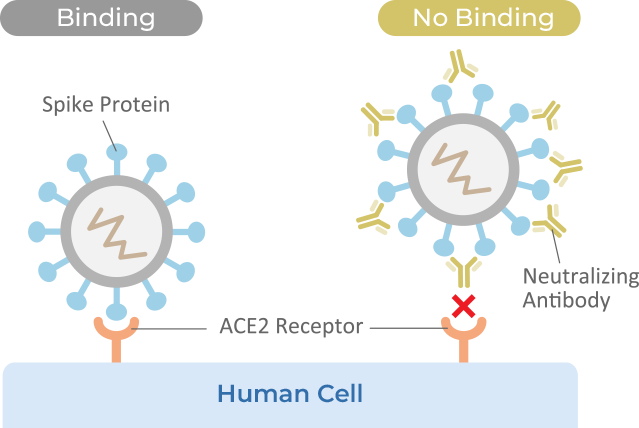
Figure 1: Principle of the Virus Neutralization Test
Fig. 1: The success of a SARS-Cov-2 virus infection is dependent on the binding of the receptor binding domain (RBD) of the virus binding to the ACE2 receptors of the human host cell (hACE2). Neutralizing antibodies, if present, bind the RBD region inhibiting the virus from binding the hACE2 receptors that then prevents the virus’ ability to infect the host cell.
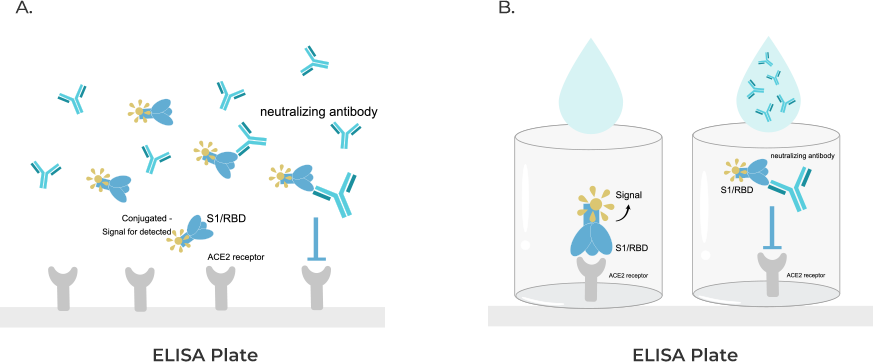
Figure 2: Applying Surrogate Virus Neutralization Technology
Fig. 2: a, In the surrogate virus neutralization test (sVNT), the anti-SARS-CoV-2 neutralizing antibodies block HRP-conjugated RBD protein from binding to the hACE2 protein pre-coated on an ELISA plate. b, Due to the nature of the blocking format of the assay, a sample without neutralizing antibody will allow the HRP-conjugated RBD to bind the ACE2 on the plate, resulting in the enzymatic color change. Alternatively, if the sample does contain neutralizing antibody, the binding and enzymatic reaction will not proceed, thus little to no color change occurs.
Real-World Application: Global Impact and Accessibility
The GenScript cPass™ SARS-CoV-2 Neutralizing Antibody Detection Kit has had a profound impact on both clinical and research settings worldwide:
1. Monitoring Immune Response
2. Widespread Adoption
3. Contributed to Wildlife Surveillance
Results
The development of GenScript’s cPass™ SARS-CoV-2 Neutralization Antibody Detection Kit exemplifies the crucial role of antibody-based diagnostics in addressing global health challenges. By creating a highly sensitive, specific, and accessible tool for detecting neutralizing antibodies, GenScript has provided an invaluable resource for clinicians, researchers, and public health officials in the battle against COVID-19. The success of this assay highlights the importance of innovation and adaptability in developing diagnostics that meet the evolving needs of a global pandemic.
In the highly competitive field of in vitro diagnostics, optimizing assay sensitivity is not just an option—it's a necessity. The ability to detect even the smallest amounts of a target biomarker can be the difference between a timely diagnosis and a missed opportunity for early intervention. As we’ve explored, the sensitivity of antibody-based immunoassays is influenced by several critical factors, including antibody affinity, specificity, and the choice of signal amplification and detection systems. By carefully balancing these elements, you can enhance the performance of your diagnostic assays, ensuring they are both reliable and clinically relevant.
The journey from developing an immunoassay to its successful application in real-world settings, as illustrated in our own case study developing GenScript’s cPass™ SARS-CoV-2 Neutralization Antibody Detection Kit, demonstrates the importance of optimizing sensitivity. Whether you are working on detecting low-abundance biomarkers in infectious diseases, cancer, or other conditions, the strategies discussed here will empower you to push the boundaries of what is possible in diagnostic accuracy.
As the demand for precision diagnostics continues to grow, the need for ultra-sensitive assays will only become more critical. By implementing the best practices outlined in this article, you can ensure that your immunoassays are not just meeting but exceeding the expectations of today’s healthcare landscape, ultimately leading to better patient outcomes and more effective disease management.
1) “In Vitro Diagnostics.” WHO, www.who.int/health-topics/in-vitro-diagnostics. Accessed 3 Sept. 2024
2) “In Vitro Diagnostics.” FDA, www.fda.gov/medical-devices/products-and-medical-procedures/in-vitro-diagnostics. Accessed 3 Sept. 2024.
3) Cox, Karen L., et al. “Immunoassay Methods.” Assay Guidance Manual - NCBI Bookshelf, 8 July 2019, www.ncbi.nlm.nih.gov/books/NBK92434.
4) IVD Raw Material Market: Global Industry Insights, Trends, Outlooks, and Opportunity Analysis, 2022-2030. Coherent Market Insights.
5) Darwish IA. Immunoassay Methods and their Applications in Pharmaceutical Analysis: Basic Methodology and Recent Advances. Int J Biomed Sci. 2006 Sep;2(3):217-35. PMID: 23674985; PMCID: PMC3614608.
6) Coronavirus disease (COVID-19) – World Health Organization. 12 July 2024, www.who.int/emergencies/diseases/novel-coronavirus-2019.
7) Tan, C.W., Chia, W.N., Qin, X. et al. A SARS-CoV-2 surrogate virus neutralization test based on antibody-mediated blockage of ACE2–spike protein–protein interaction. Nat Biotechnol 38(9), 1073–1078 (2020). https://doi.org/10.1038/s41587-020-0631-z
8) cPass SARS-CoV-2 Neutralization Antibody Detection Kit. 1 Feb. 2022, www.genscript.com/gsfiles/techfiles/EUA%2dGenScript%2dcpass%2difu.pdf?=20220217?1283406859.
9) Wanchaijiraboon, Passakorn, et al. “Durability of Immune Response to ChAdOx1-nCoV-19 Vaccine in Solid Cancer Patients Undergoing Anticancer Treatment.” Vaccines, vol. 10, no. 10, Oct. 2022, p. 1662, doi:10.3390/vaccines10101662.
10) Chandler, Jeffrey C., et al. “SARS-CoV-2 exposure in wild white-tailed deer (Odocoileus virginianus).” Proceedings of the National Academy of Sciences, vol. 118, no. 47, Nov. 2021, doi:10.1073/pnas.2114828118.
11) Vandegrift, Kurt J., et al. “SARS-CoV-2 Omicron (B.1.1.529) Infection of Wild White-Tailed Deer in New York City.” Viruses, vol. 14, no. 12, Dec. 2022, p. 2770, doi:10.3390/v14122770.