- REAGENT SERVICES Hot!
-
PRODUCTS
-
 Most Popular Reagents
-
 Instruments
-
 Antibodies
-
ELISA Kits
-
 Protein Electrophoresis and Blotting
-
Protein and Antibody Purification
-
Recombinant Proteins
-
 Molecular Biology
-
 Stable Cell Lines
-
 Cell Isolation and Activation
-
 IVD Raw Materials
-
 Therapy Applications
-
 Resources
-
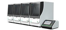
![AmMag™ Quatro Automated Plasmid Purification]() AmMag™ Quatro automated plasmid purification
AmMag™ Quatro automated plasmid purification
-
![Anti-Camelid VHH]() MonoRab™ Anti-VHH Antibodies
MonoRab™ Anti-VHH Antibodies
-
![ELISA Kits]() ELISA Kits
ELISA Kits
-
![Precast Gels]() SurePAGE™ Precast Gels
SurePAGE™ Precast Gels
-
![Quatro ProAb Automated Protein and Antibody Purification System]() AmMag™ Quatro ProAb Automated Protein and Antibody Purification System
AmMag™ Quatro ProAb Automated Protein and Antibody Purification System
-
![Target Proteins]() Target Proteins
Target Proteins
-
![AmMag™ Quatro Automated Plasmid Purification]() AmMag™ Quatro automated plasmid purification
AmMag™ Quatro automated plasmid purification
-
![Stable Cell Lines]() Stable Cell Lines
Stable Cell Lines
-
![Cell Isolation and Activation]() Cell Isolation and Activation
Cell Isolation and Activation
-
![Quick
Order]() Quick Order
Quick Order
-
![Quick
Order]() Quick Order
Quick Order
- APPLICATIONS
- RESOURCES
- ABOUT US


DNA visualization is an important application in understanding a variety of cellular processes, such as replication, transcription, and recombination, and the interactions between DNA and associated proteins and RNA. Two techniques are commonly used for DNA imaging, fluorescence in situ hybridization (FISH) and fluorescent tagging of DNA-binding proteins. FISH uses fluorescently tagged nucleic acid probes to bind and visualize DNA. While this technique offers the flexibility to target specific sequences through base pairing of the nucleic acid probes, it cannot be used for live imaging because of the requirement for sample fixation. Conversely, proteins tagged with a fluorescent label can be used for live imaging, but are limited by their fixed target sequences, restricting their use mostly to repetitive DNA sequences, such as telomeres.
New advances in CRISPR/Cas9 technology offer the benefits of both live imaging and easy target sequence customization and flexibility. Inactivated dCas9 can be tagged with fluorophores for imaging both repetitive DNA elements and protein-encoding genes, enabling us to observe chromatin organization throughout the cell cycle. In addition to live DNA imaging, the CRISPR/Cas9 system can be used for live RNA imaging as well. Modifications to the gRNA sequence allow for mRNA recognition and tracking. Using CRISPR-mediated RNA imaging techniques, researchers have been able to visualize the accumulation of ACTB, CCNA2 and TRFC mRNAs in RNA granules. These new applications improve existing methodologies for live imaging within cells allowing for the study of dynamic cellular processes involving DNA and RNA.
CRISPR Services
 CRISPR gRNA Plasmids
Licensed from the Feng Zhang Laboratory at the Broad Institute
Learn More CRISPR
sgRNAA powerful tool for creating gene knock-ins and knock-outs
Learn More HDR Knock-in Templates
ssDNA and dsDNA HDR donor templates for effective gene knock-in
Learn More CRISPR Cell Lines
Genetically modified cell line in any mammalian cell line and targeting any gene.
Learn More Microbial Gene Editing
Highly efficient, seamless genome editing in E.coli using CRISPR technology
Learn More CRISPR gRNA Libraries
Genome-wide gRNA libraries for efficient screening
Learn More -