- REAGENT SERVICES Hot!
-
PRODUCTS
-
 Most Popular Reagents
-
 Instruments
-
 Antibodies
-
ELISA Kits
-
 Protein Electrophoresis and Blotting
-
Protein and Antibody Purification
-
Recombinant Proteins
-
 Molecular Biology
-
 Stable Cell Lines
-
 Cell Isolation and Activation
-
 IVD Raw Materials
-
 Therapy Applications
-
 Resources
-
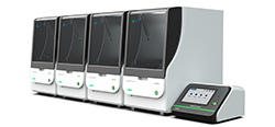
![AmMag™ Quatro Automated Plasmid Purification]() AmMag™ Quatro automated plasmid purification
AmMag™ Quatro automated plasmid purification
-
![Anti-Camelid VHH]() MonoRab™ Anti-VHH Antibodies
MonoRab™ Anti-VHH Antibodies
-
![ELISA Kits]() ELISA Kits
ELISA Kits
-
![Precast Gels]() SurePAGE™ Precast Gels
SurePAGE™ Precast Gels
-
![Quatro ProAb Automated Protein and Antibody Purification System]() AmMag™ Quatro ProAb Automated Protein and Antibody Purification System
AmMag™ Quatro ProAb Automated Protein and Antibody Purification System
-
![Target Proteins]() Target Proteins
Target Proteins
-
![AmMag™ Quatro Automated Plasmid Purification]() AmMag™ Quatro automated plasmid purification
AmMag™ Quatro automated plasmid purification
-
![Stable Cell Lines]() Stable Cell Lines
Stable Cell Lines
-
![Cell Isolation and Activation]() Cell Isolation and Activation
Cell Isolation and Activation
-
![Quick
Order]() Quick Order
Quick Order
-
![Quick
Order]() Quick Order
Quick Order
- APPLICATIONS
- RESOURCES
- ABOUT US


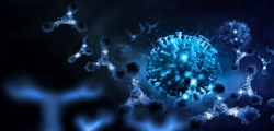
New Opportunities with a Neoantigen-Based Strategy in Rare Liver Cancer
What is fibrolamellar carcinoma?
Fibrolamellar carcinoma (FLC) is a rare form of liver carcinoma that affects young and otherwise healthy individuals. It’s insidious in that it does not present overt symptoms early on, often spreading to other organs and tissues before diagnosis.1 Driving this rare form of hepatocellular carcinoma is a common denominator, a fusion protein frequently found within the tumor and first identified in 2014 by a team at Rockefeller University.2
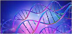
The chimeric transcript was discovered and consistently detected through RNA sequencing analysis of tumor tissue from 15 patients. It was shown to result from the fusion of coding sequences from DNAJB1, a heat shock 40 protein family member, and PRKACA, the cAMP-dependent protein kinase A (PKA) catalytic subunit alpha.2 The resulting DNAJ-PKAc fused protein is functional, conserving PKA’s kinase activity, and is a well-established marker and driver of FLC.

FLC driver mutation results from a 400 kb deletion in chromosome 19 and the consequent in-frame fusion of DNAJB1 exon1, and PRKACA exons 2-10. The resulting protein chimera with conserved kinase activity is proposed to drive the tumorigenesis process.3 Retrieved without modifications from “What is Fibrolamellar carcinoma,” What is Fibrolamellar Carcinoma? - Fibrolamellar Cancer Foundation (fibrofoundation.org)>1, 2
DNAJ-PKAc: Failed strategies against a complex oncogenic driver
In FLC, the DNAJ-PKAc fusion protein appears to contribute to tumor progression and treatment resistance via different mechanisms, including aberrant kinase activity, abnormal protein-protein interactions, and upregulation of oncogenic signaling pathways. For instance, protein and transcript analysis have shown several upregulated signaling pathways in FLC, such as aromatase and aurora A kinase, which are known oncogenic pathways in other cancer types.4,5 Most recently, DNAJ-PKAc has been shown to interact with an atypical binder, Bcl-2-associated athanogene 2 (BAG2), which is upregulated in FLC. Significantly, the elucidated DNAJ-PKAc /Hsp70/BAG2/Bcl-2 axis is linked to chemotherapy resistance through anti-apoptotic signaling.6
Unfortunately, to date, studies targeting aberrant kinase activity or specific signaling nodes downstream of DNAJ-PKAc have not been fruitful, showing either high toxicity preclinically or poor responses in FLC patients, respectively.4,5
A new approach leverages DNAJ-PKAc fusion neoantigen
Because of the lack of effective therapies, FLC patients continue to rely primarily on surgical tumor resection, which is only successful in the absence of metastasis and has a survival rate of 30% to 45% at five years.2 Following metastasis, FLC is not responsive to other liver cancer treatments.
Immunotherapy has emerged as an effective strategy to redirect and strengthen the immune system against cancer cells. Whether immunotherapy strategies are effective for FLC is unknown. However, several studies indicate that FLC patients could benefit from strengthened T-cell immunity. Recently a team at the Department of Immunology, St. Jude Children’s Research Hospital, led by Dr. Paul G Thomas, has explored the potential of leveraging DNAJ-PKA fusion antigens for immunotherapy in FLC patients.5
Identifying immunogenic neoepitopes
Not all tumor-derived antigens or neoantigens are immunogenic. The identification of immunogenic tumor-specific antigens is a critical step in developing cancer vaccines and T-cell therapies. Commonly in silico predictions help prioritize neoepitopes based on their MHC binding.
Therefore, as a first step, the team evaluated whether DNAJ-PKA-derived fusion peptides (i.e., 8-15 mer peptides spanning the fusion sequence) were presented via MHC molecules. Based on their analysis, they predicted the binding of various DNAJ-PKA neoepitopes to both class I and class II HLAs, including HLA alleles expressed by a cohort of FLC patients. Because HLA I binders were more frequent, the team leveraged HLA I multimer reagents, representing alleles expressed by FLC patients for biochemical binding assays, uncovering over 30 neoepitope binders. Significantly, identified fusion neoepitope binders could be matched to some of those predicted in most FLC patients, demonstrating their potential utility as immunogens.
“The corresponding peptides (Table S2) were commercially synthesized (Genscript) and diluted to 1 mM in the manufacturer-recommended solvent (ddH2O or DMSO, depending on solubility).”
The Thomas group then developed HLA I monoallelic antigen-presenting cells to further validate binding. One of the alleles expressed, HLA-A∗68:02, was predicted to bind various fusion peptides. By co-expressing this allele and the DNAJB1-PRKACA fusion in antigen-presenting cells, they validated the presentation of the specific fusion peptide, EIFDRYGEEV.
“Full length coding sequences for these alleles were obtained from the IPD-IMGT/HLA database,then synthesized and cloned via restriction sites (Genscript) into lentiviral vector pLVX-EF1α-IRES-Puro”
However, the presentation of a neoepitope does not guarantee its immunogenicity. Therefore, once they honed on this interaction, the group inquired about patient-derived T-cell responses. When tumor-infiltrating T cells from the HLA I allele-matched patient were stimulated by the antigen-presenting cells (i.e., HLA-A∗68:02/ EIFDRYGEEV), the team confirmed robust responses, including production of IFNγ and TNFα.
Conclusion
The finding of immune reactivity to DNAJ-PKA-derived fusion peptides opens up the possibility of developing an effective immunotherapy for FLC patients. The Thomas group went further to perform single T-cell analyses and identified various TCR sequences with reactivity to the fusion neoantigen EIFDRYGEEV. A similar approach implemented across various patients enabled them to initially identify over 20 DNAJ-PKA fusion peptide-specific CD8+ TCRs. Despite the numerous candidates, further validation significantly reduced the total of truly reactive TCRs, highlighting the challenges of identifying responses to DNAJ-PKA-derived fusion peptides, which may be rare. A silver lining in their findings is that despite their rarity, T cell responses to DNAJ-PKA- fusion peptides can be productive, directing specific cytotoxicity.
Reference
[1] Fibrolamellar Cancer Foundation (n.d.). What is Fibrolamellar? Https://Fibrofoundation.org/. https://fibrofoundation.org/about-fibro/what-is-fibrolamellar-carcinoma/
[2] Honeyman JN, et al. (2014), Detection of a recurrent DNAJB1-PRKACA chimeric transcript in fibrolamellar hepatocellular carcinoma. Science. https://doi.org/10.1126/science.1249484
[3] Riggle KM, et al. (2016), Fibrolamellar Hepatocellular Carcinoma: Mechanistic Distinction From Adult Hepatocellular Carcinoma. Pediatr Blood Cancer. https://doi.org/10.1002/pbc.25970
[4] Lalazar G, Simon SM. (2018), Fibrolamellar Carcinoma: Recent Advances and Unresolved Questions on the Molecular Mechanisms. Semin Liver Dis. https://doi.org/10.1055/s-0037-1621710
[5] Kirk AM, et al. (2024), DNAJB1-PRKACA fusion neoantigens elicit rare endogenous T cell responses that potentiate cell therapy for fibrolamellar carcinoma. Cell Rep Med. https://doi.org/10.1016/j.xcrm.2024.101469
[6] Lauer SM, et al. (2024), Recruitment of BAG2 to DNAJ-PKAc scaffolds promotes cell survival and resistance to drug-induced apoptosis in fibrolamellar carcinoma. Cell Rep. https://doi.org/10.1016/j.celrep.2024.113678
-