-
REAGENT SERVICES
Hot!
-
Most Popular Services
-
Molecular Biology
-
Recombinant Antibody/Protein
-
Reagent Antibody
-
CRISPR Gene Editing
-
DNA Mutant Library
-
IVT RNA and LNP Formulations
-
Oligo Synthesis
-
Peptides
-
Cell Engineering
-
- Gene Synthesis FLASH Gene
- GenBrick™ Up to 200kb
- Gene Fragments Up to 3kb now
- Plasmid DNA Preparation Upgraded
- Cloning and Subcloning
- ORF cDNA Clones
- mRNA Plasmid Solutions New!
- Cell free mRNA Template New!
- AAV Plasmid Solutions New!
- Mutagenesis
- GenCircle™ Double-Stranded DNA New!
- GenSmart™ Online Tools
-
-
PRODUCTS
-
Most Popular Reagents
-
 Instruments
-
Antibodies
-
ELISA Kits
-
Protein Electrophoresis and Blotting
-
Protein and Antibody Purification
-
Recombinant Proteins
-
Molecular Biology
-
Stable Cell Lines
-
Cell Isolation and Activation
-
 IVD Raw Materials
-
 Therapy Applications
-
Resources
-
- All Instruments
- Automated Protein and Antibody Purification SystemNew!
- Automated Plasmid MaxiprepHot!
- Automated Plasmid/Protein/Antibody Mini-scale Purification
- eBlot™ Protein Transfer System
- eStain™ Protein Staining System
- eZwest™ Lite Automated Western Blotting Device
- CytoSinct™ 1000 Cell Isolation Instrument
-
- Pharmacokinetics and Immunogenicity ELISA Kits
- Viral Titration QC ELISA Kits
- -- Lentivirus Titer p24 ELISA KitHot!
- -- MuLV Titer p30 ELISA KitNew!
- -- AAV2 and AAVX Titer Capsid ELISA Kits
- Residual Detection ELISA Kits
- -- T7 RNA Polymerase ELISA KitNew!
- -- BSA ELISA Kit, 2G
- -- Cas9 ELISA KitHot!
- -- Protein A ELISA KitHot!
- -- His tagged protein detection & purification
- dsRNA ELISA Kit
- Endonuclease ELISA Kit
- COVID-19 Detection cPass™ Technology Kits
-
- Automated Maxi-Plasmid PurificationHot!
- Automated Mini-Plasmid PurificationNew!
- PCR Reagents
- S.marcescens Nuclease Benz-Neburase™
- DNA Assembly GenBuilder™
- Cas9 / Cas12a / Cas13a Nucleases
- Base and Prime Editing Nucleases
- GMP Cas9 Nucleases
- CRISPR sgRNA Synthesis
- HDR Knock-in Template
- CRISPR Gene Editing Kits and Antibodies
-
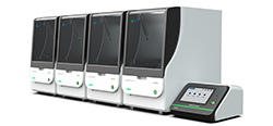
![AmMag™ Quatro Automated Plasmid Purification]() AmMag™ Quatro automated plasmid purification
AmMag™ Quatro automated plasmid purification
-
![Anti-Camelid VHH]() MonoRab™ Anti-VHH Antibodies
MonoRab™ Anti-VHH Antibodies
-
![ELISA Kits]() ELISA Kits
ELISA Kits
-
![Precast Gels]() SurePAGE™ Precast Gels
SurePAGE™ Precast Gels
-
![Quatro ProAb Automated Protein and Antibody Purification System]() AmMag™ Quatro ProAb Automated Protein and Antibody Purification System
AmMag™ Quatro ProAb Automated Protein and Antibody Purification System
-
![Target Proteins]() Target Proteins
Target Proteins
-
![AmMag™ Quatro Automated Plasmid Purification]() AmMag™ Quatro automated plasmid purification
AmMag™ Quatro automated plasmid purification
-
![Stable Cell Lines]() Stable Cell Lines
Stable Cell Lines
-
![Cell Isolation and Activation]() Cell Isolation and Activation
Cell Isolation and Activation
-
 IVD Raw Materials
-
![Quick
Order]() Quick Order
Quick Order
-
![Quick
Order]() Quick Order
Quick Order
- APPLICATIONS
- RESOURCES
- ABOUT US
- SIGN IN My Account SIGN OUT
- REGISTER

![Immunoprecipitation Immunoprecipitation]()
Immunoprecipitation
Resources » Research Applications » Immunoprecipitation
Immunoprecipitation (IP) is a technique to isolate a specific protein out of a solution using an antibody that binds to it. Antibody-protein complexes are removed from the solution with the addition of an insoluble form of an antibody binding protein, such as Protein A or Protein G conjugated to agarose slurry or the newly popular Magnetic Beads. Immunoprecipitation assays detect the interaction of a target protein with other proteins or nucleic acids. Assay examples include Co-IP, ChIP, RIP and tagged protein IP.

Co-Immunoprecipitation (Co-IP) is a powerful method that is most widely used by researchers to analyze protein–protein interactions. This process provides a rapid and simple method to separate a specific protein from a sample containing thousands of different proteins, such as serum, cell lysate, homogenized tissue or conditioned media.
Chromatin Immunoprecipitation (ChIP) is a type of immunoprecipitation used to investigate regions of genome associated with a target DNA-binding protein, or conversely to identify specific proteins associated with a particular region of the genome. The primary applications of ChIP are as follows:
- Binding site consensus identification
- Target gene identification
- Analysis of epigenetic phenomenon
RNA Immunoprecipitation (RIP) is performed with an antibody that targets a specific RNA-binding protein. By isolating the protein, the RNA bound to it is also isolated. The RNA-protein complexes are separated by RNA extraction. The RNA can be analyzed by cDNA sequencing or RT-PCR.
Procedure
Traditional immunoprecipitation (IP) uses agarose beads coated in protein A/G. Most of these IP protocols require more than three washing steps to eliminate background and non-specific binding. Each step of washing causes some bead loss or leaves behind residual contaminants.
The use of magnetic beads has recently gained popularity as a quicker, more accurate approach for immunoprecipitation. Unlike agarose, no columns or centrifugations are required. Also, you can significantly reduce experiment variability by being able to remove all supernatant without disturbing your pellet. You will consistently get more reliable and reproducible data with GenScript MagBeads.
This protocol offers a general guideline for immunoprecipitation with GenScript MagBeads.
A. Cross-linking IgG to the Beads
- Add 1 ml 0.2 M triethanolamine, pH8.2 to the Protein A/G MagBeads with immobilised IgG. Wash twice using the magnetic separation rack with 0.2 M triethanolamine, pH8.2 as the washing buffer.
- Resuspend the beads in 1 ml of 20 mM dimetyl pimelimidate dihydrochloride (DMP) in 0.2 M triethanolamine, pH8.2 (5.4 mg DMP/ml buffer). This cross-linking solution must be prepared fresh.
- Incubate the beads with rotational mixing for 30 minutes at room temperature. Use the magnetic separation rack to collect the beads and discard the supernatant.
- Resuspend the beads in 1 ml of 50 mM Tris, pH7.5 to stop the reaction and incubate for 15 minutes at room temperature with rotational mixing.
- Use the magnetic separation rack to collect the beads and discard the supernatant. Wash the cross-linked beads three times with 1 ml PBS, pH7.4.
B. Binding Target Protein to the IgG Cross-linked Beads
- Add sample containing target protein to the beads. For a 100 kDa protein, use a volume containing approximately 25 µg of target protein/ml beads to ensure an excess of protein. If dilution of target protein is necessary, PBS or 0.1 M phosphate buffer (pH7-8) can be used as the dilution buffer.
- Incubate with tilting and rotation for one hour at room temperature.
- Place the tube on the magnetic separation rack for 2 minutes to collect the IgG‐coated beads‐target protein complex. For viscous samples, double the time on the rack. Discard the supernatant.
- Wash the beads 3 times using 1 ml PBS.
C. Elution of Target Protein
-
Denaturing elution
Place the tube on the magnetic separation rack to collect the beads and discard the supernatant. Add 100 µl 1XSDS Sample Buffer to the tube and mix well. Heat the tube at 100°C for five minutes. Use the magnetic separation rack to collect the beads and transfer the supernatant containing desired sample into a new tube. Analyze the sample by SDS-PAGE followed by Western blot analysis. -
Non‐denaturing elution
Place the tube on the magnetic separation rack to collect the beads and discard the supernatant. Add 100 µl Elution Buffer to the tube and mix well. Incubate for five minutes at room temperature with occasional mixing. Use the magnetic separation rack to collect the beads and transfer the supernatant into a new tube. Repeat the elution and incubation step twice. Add 10 μl Neutralization Buffer to each 100 μl of eluation buffer to neutralize the pH.
Related Products
Immunoprecipitation Products
Protein A, G and A/G MagBeadsimmunoprecipitation and micro-scale antibody purification.
Learn MoreNi-charged and Glutathione MagBeadsmicro-scale purification of His and GST tagged proteins.
Learn More
-


