Fluorescent Modifications for Peptides
GenScript offers numerous fluorescent tags for peptides, and our
repertoire is being expanded continuously. Listed below are a few
of our most commonly used modifications:
| Name |
Excitation (nm) |
Emission (nm) |
Emission color |
Application Fields |
|
7-Methoxycoumarin-4-acetic acid
|
328
|
393
|
Blue
|
In vitro imaging
Subcellular localization
Confocal microscopy
Flow cytometry
|
|
FITC-Ahx
|
494
|
521
|
Green
|
|
FAM
|
495
|
520
|
Green
|
|
Cy3
|
555
|
570
|
Yellow
|
|
5-Carboxytetramethylrhodamine (TMR)
|
542
|
568
|
Orange
|
|
Cy5
|
646
|
662
|
Red
|
|
Cy5.5
|
673
|
707
|
Near-infrared
|
In vitro imaging
Subcellular localization
In vivo optical imaging
Angiography
|
|
Cy7
|
750
|
773
|
Near-infrared
|
FRET pairs
Fluorescence resonance energy transfer (FRET) is a mechanism that
describes the energy transfer between two fluorophores. Since FRET
efficiency is partly based on distance between a donor and
acceptor molecule, this technique is commonly used for studying
enzyme efficiency, protein-protein interactions, or other
molecular dynamics (Fig 1).
Fig 1. FRET mechanism for protease studies. When the
peptide remains intact, the acceptor molecule will quench the
donor molecule, and no fluorescence will be detected. If the
sequence is cleaved by protease activity, the acceptor will no
longer quench the donor, and a fluorescent signal will be
detected.
Below are the most commonly used FRET pairs:
| Donor |
Acceptor |
|
Name
|
Excitation (nm)
|
Emission (nm)
|
Name
|
Excitation (nm)
|
Emission (nm)
|
|
Cy2
|
490
|
510
|
Cy3
|
555
|
570
|
|
FITC
|
494
|
521
|
TRITC
|
557
|
576
|
|
FAM
|
495
|
520
|
Cy3
|
555
|
570
|
|
FAM
|
495
|
520
|
Texas Red
|
589
|
615
|
|
FAM
|
495
|
520
|
Cy5
|
646
|
662
|
|
Cy3
|
555
|
570
|
Cy5
|
646
|
662
|
|
EDANS
|
335
|
493
|
DABCYL
|
453
|
-
|
|
Glu(EDANS)-NH2
|
335
|
493
|
DABCYL
|
453
|
-
|
|
MCA
|
328
|
393
|
DNP
|
348
|
-
|
|
Abz
|
330
|
420
|
DNP
|
348
|
-
|
|
Abz
|
330
|
420
|
Tyr (3-NO2)
|
360
|
-
|
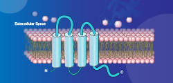
Fluorescent peptides for subcellular imaging
In vitro imaging by confocal or fluorescent microscopy remains one
of the most efficient and effective methods for studying a variety
of biological process and interactions in cells. When combined
with imaging, fluorescent-labelled peptides can be used to
identify specific targets.
Read More »
Fluorescent peptides for angiography
In vivo imaging for angiography continues to be a major
application for fluorescent-labeled peptides. This technique, in
which contrast agents are used to image the inside of blood
vessels, enables physicians to choose the most appropriate medical
strategies or treatments for an individual.
Read More »
FRET for screening proteolytic peptides
Proteolytic enzymes play important roles in infectious diseases,
which makes them a target for developing novel therapeutics. To
identify the peptide sequences that these enzymes target, peptide
libraries are often used. The potential protease target sequences
are combined with FRET pairs, such that when the enzyme cleaves
the target peptide, a fluorescent signal can be detected.
Read More »
References
Fluorescent Modification Case studies
Fluorescent peptide labels have numerous research applications,
and GenScript has extensive experience synthesizing peptides with
a variety of modifications.
Case Study 1
Sequence: LYRLGLGH
Modification: MCA/DNP
Quantity: 1-4 mg
| Required purity |
Estimated Turnaround time |
Actual purity |
Actual turnaround time |
|
>98%
|
17 days
|
99.563%
|
14 days
|
Click here to view HPLC results »
Click here to view
MS results »
Case Study 2
Sequence: IKDLSKEERLWEVQRILTALKRKLREA
Modification: 5-FAM (N-terminal)
Quantity: 10-14 mg
| Required purity |
Estimated Turnaround time |
Actual purity |
Actual turnaround time |
|
>98%
|
23 days
|
99.10%
|
13 days
|
Click here to view
HPLC results »
Click here to view
MS results »
Case Study 3
Sequence: RAKWNNTLKQIASK
Modification: FITC-Ahx (N-terminal)
Quantity: 5-9 mg
| Required purity |
Estimated Turnaround time |
Actual purity |
Actual turnaround time |
|
>98%
|
17 days
|
99.81%
|
5 days
|
Click here to view
HPLC results »
Click here to view
MS results »
 Most Popular Products
 CRISPR Gene Editing
 Antibodies
 Protein Electrophoresis and Blotting
 Molecular Biology
 Stable Cell Lines
 Cell Therapy
 Diagnostics
 Resources


 next to the
desired modification or indicate the modification in the comments
section on the secure
next to the
desired modification or indicate the modification in the comments
section on the secure