- REAGENT SERVICES Hot!
-
PRODUCTS
-
 Most Popular Products
-
 CRISPR Gene Editing
-
 Antibodies
-
ELISA Kits
-
 Protein Electrophoresis and Blotting
-
Protein Purification
-
Proteins
-
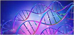
 Molecular Biology
-
 Stable Cell Lines
-
 Cell Therapy
-
 Diagnostics
-
 Resources
-
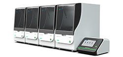
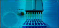
![AmMag™ Quatro Automated Plasmid Purification]() AmMag™ Quatro automated plasmid purification
AmMag™ Quatro automated plasmid purification
-
![Cas
Nucleases]() Cas Nucleases
Cas Nucleases
-
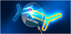
![Anti-Camelid
VHH]() MonoRab™ Anti-VHH Antibodies
MonoRab™ Anti-VHH Antibodies
-
![AAV2 and AAVX Titer Capsid ELISA Kits]() AAV2 and AAVX Titer Capsid ELISA Kits
AAV2 and AAVX Titer Capsid ELISA Kits
-
![Precast Gels]() Precast Gels
Precast Gels
-
![Protein
Isolation &
Purification]() Protein Isolation & Purification
Protein Isolation & Purification
-
![Recombinant Cytokines]() Recombinant Cytokines
Recombinant Cytokines
-
![AmMag™ Quatro Automated Plasmid Purification]() AmMag™ Quatro Customer Testimonial
AmMag™ Quatro Customer Testimonial
-
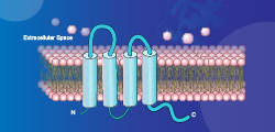
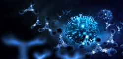
![Claudin 18.2]() Claudin 18.2
Claudin 18.2
-
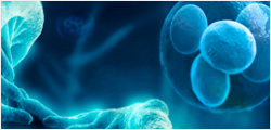
![Cell Isolation]() Cell Therapy Comprehensive Product Solutions
Cell Therapy Comprehensive Product Solutions
-
![Quick
Order]() Quick Order
Quick Order
-
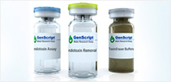
![Endotoxin
Detection &
Removal
System]() Endotoxin Detection & Removal System
Endotoxin Detection & Removal System
-
- APPLICATIONS
- RESOURCES
- ABOUT US


 Dr. Travis Thomson